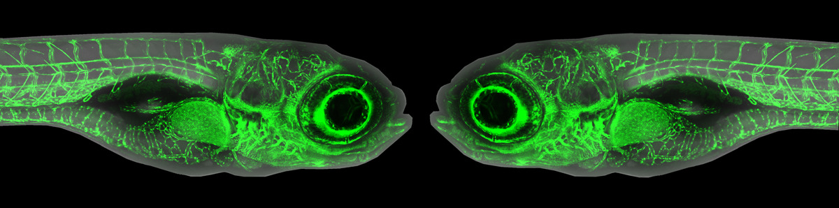
Zebrafish Images - Hematopoetic site in the Developing Zebrafish
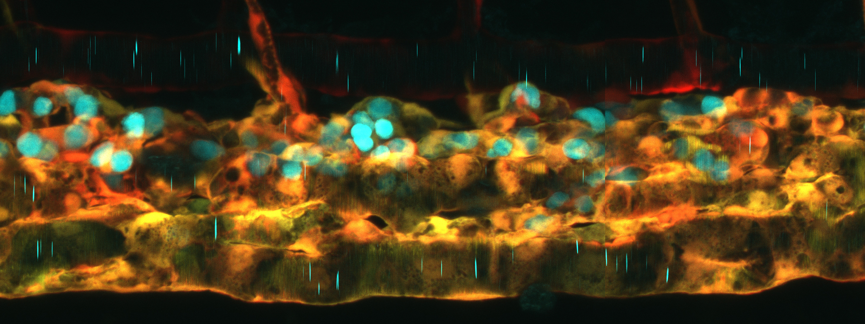
Photo Citation: Hematopoetic site in the Developing Zebrafish
Image by Dr. Laura Gutierrez-Miranda
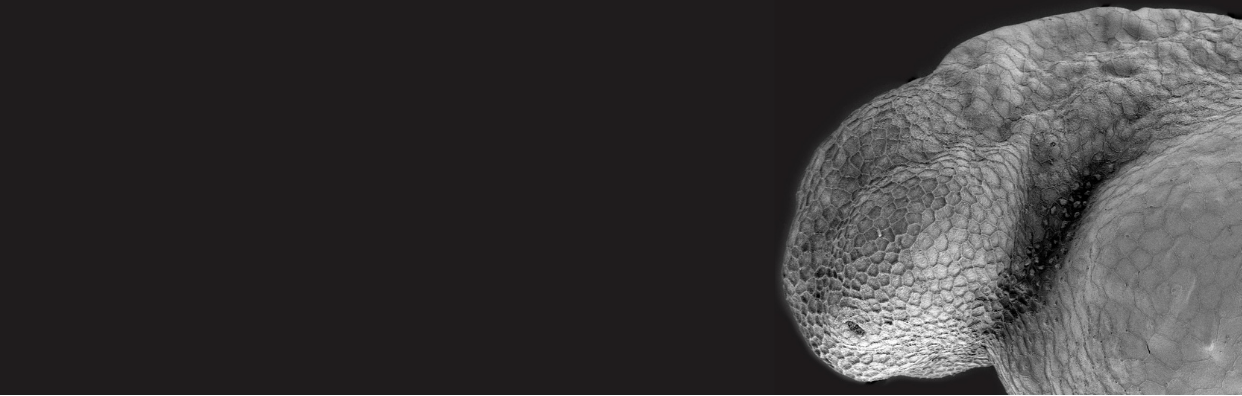
As our members may know, a more recent development in the International Zebrafish Website is to incorporate photos that convey the fascinating science being done using zebrafish as a model system for investigation and teaching, as well as celebrating the beauty of zebrafish. We are pleased to present our top pick for the next edition of the IZFS website banner: a high resolution and high-definition image captured by Laura Gutierrez Miranda. Originally hailing from Spain, Laura took this image while she was a postdoc in Karina Yaniv’s laboratory in Israel (Weizmann Institute of Science) when she was researching the development and formation of the vascular epithelium. She tells us that this photo was captured on a hot summer day in August. Because everyone else was enjoying the sunshine, Laura had some much-needed extra time to spend on the microscope that is usually fully booked. This time allowed her to play around with the laser settings capturing this crystal-clear image of the primary hematopoetic site in a 3-day post-fertilization zebrafish. To visualize the different cell types, she used the Tg(kdrl:mCherry) and Tg(runx1:EGFP) reporter lines where kdrl is expressed in the vascular epithelium (in orange) and runx1 is expressed in the caudal hematopoietic tissue (in cyan). Laura tells us that she was experimenting with a more colorblind-friendly palette, modifying the lookup tables (LUTs) so that the endothelial cells are orange and the hematopoietic stem cells are cyan, something we should all remember when collecting color images!
Since taking this image Laura has moved to Sweden where she works as postdoc in Kaska Koltowska’s Vascular Biology Laboratory at Uppsala University. There, she is exploring transcriptional regulation underlying the specification of lymphatic tissue.
We invite all IZFS members to submit photos, drawings related to zebrafish research and teaching. Submissions are ongoing and the photo selections occur three times a year. All photos should have a minimum resolution of 300 dpi with a composition oriented to the horizontal banner format seen on the website. Photo submissions should be directed to: Olivia Flood; oflood@izfs.org
Related publication: Laura Gutierrez-Miranda and Karina Yaniv (2020). Cellular Origins of the Lymphatic Endothelium: Implications for Cancer Lymphangiogenesis. Frontiers in Physiology www.frontiersin.org, Review, doi: 10.3389/fphys.2020.577584
Megan Leask, Lecturer, Dept. Of Physiology, University of Otago, New Zealand.
Kate Whitlock, Professor, Dept. Of Neuroscience, University of Valparaiso, Chile.